Background
We had previously shown that co-ingesting perilla oil with γ-cyclodextrin (γ-CD) improves the absorption of α-linolenic acid (ALA) into plasma. In this study, we used mice to examine whether this effect extends beyond plasma to the liver and red blood cell membranes as well. Since the diet contained no EPA, any increase in EPA observed in blood or tissues reflects activation of the in vivo ALA→EPA conversion pathway (the Δ6 desaturase pathway).
Four groups were established: Control (standard purified diet), CD (dietary fiber replaced with γ-CD), LP (part of the soybean oil replaced with perilla oil), and IC (all dietary fiber and part of the soybean oil replaced with powdered perilla oil complex).
From Absorption to Utilization
We traced how ALA derived from perilla oil is utilized in the body, following the pathway plasma → liver → red blood cell membranes.
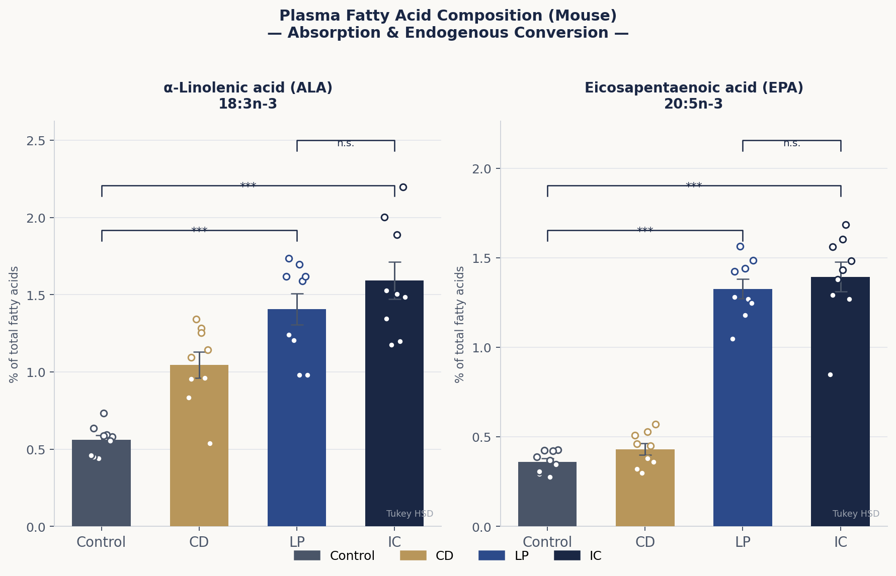
- Plasma (absorption and conversion): Both the LP and IC groups showed significantly higher ALA and EPA than the Control and CD groups (p<0.001). No significant difference was found between LP and IC (n.s.).
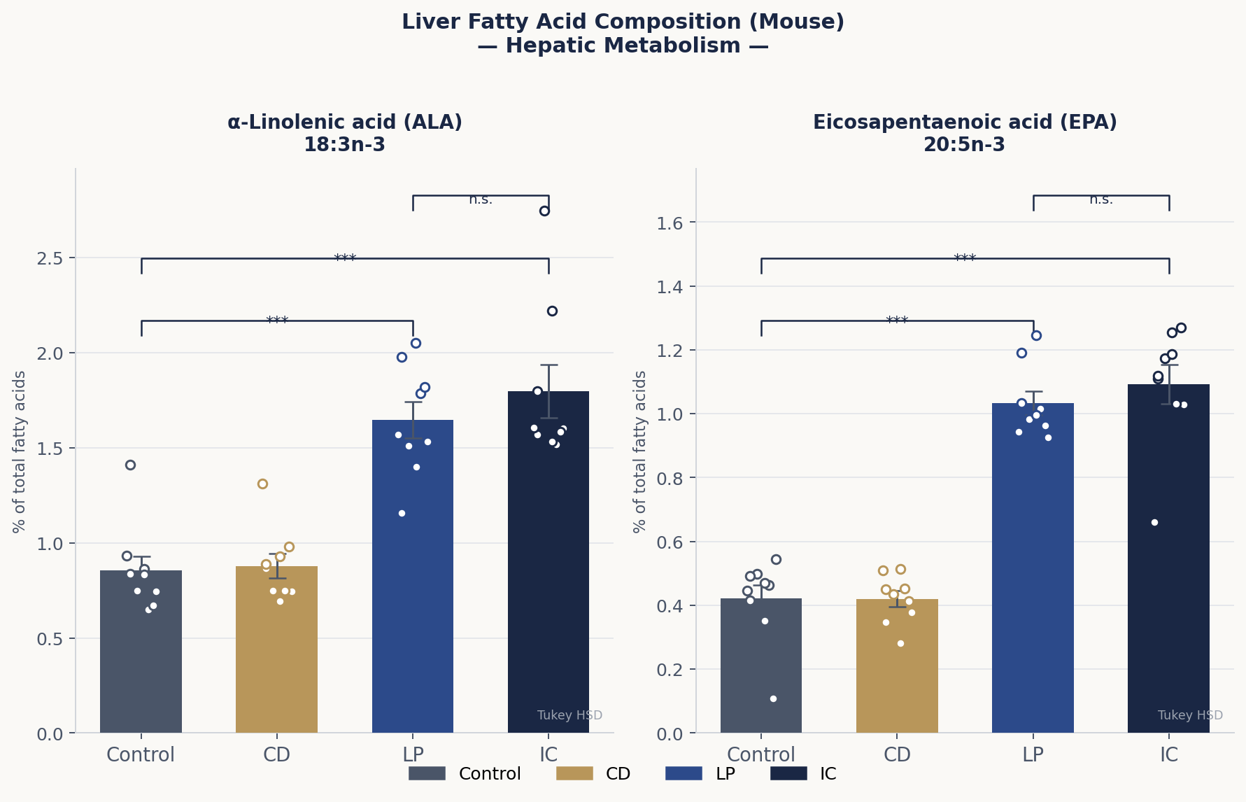
- Liver (metabolism): ALA and EPA were significantly elevated in the LP and IC groups (p<0.001). The liver, rich in Δ6 desaturase, is the primary organ for ALA→EPA conversion. No significant difference was found between LP and IC (n.s.).
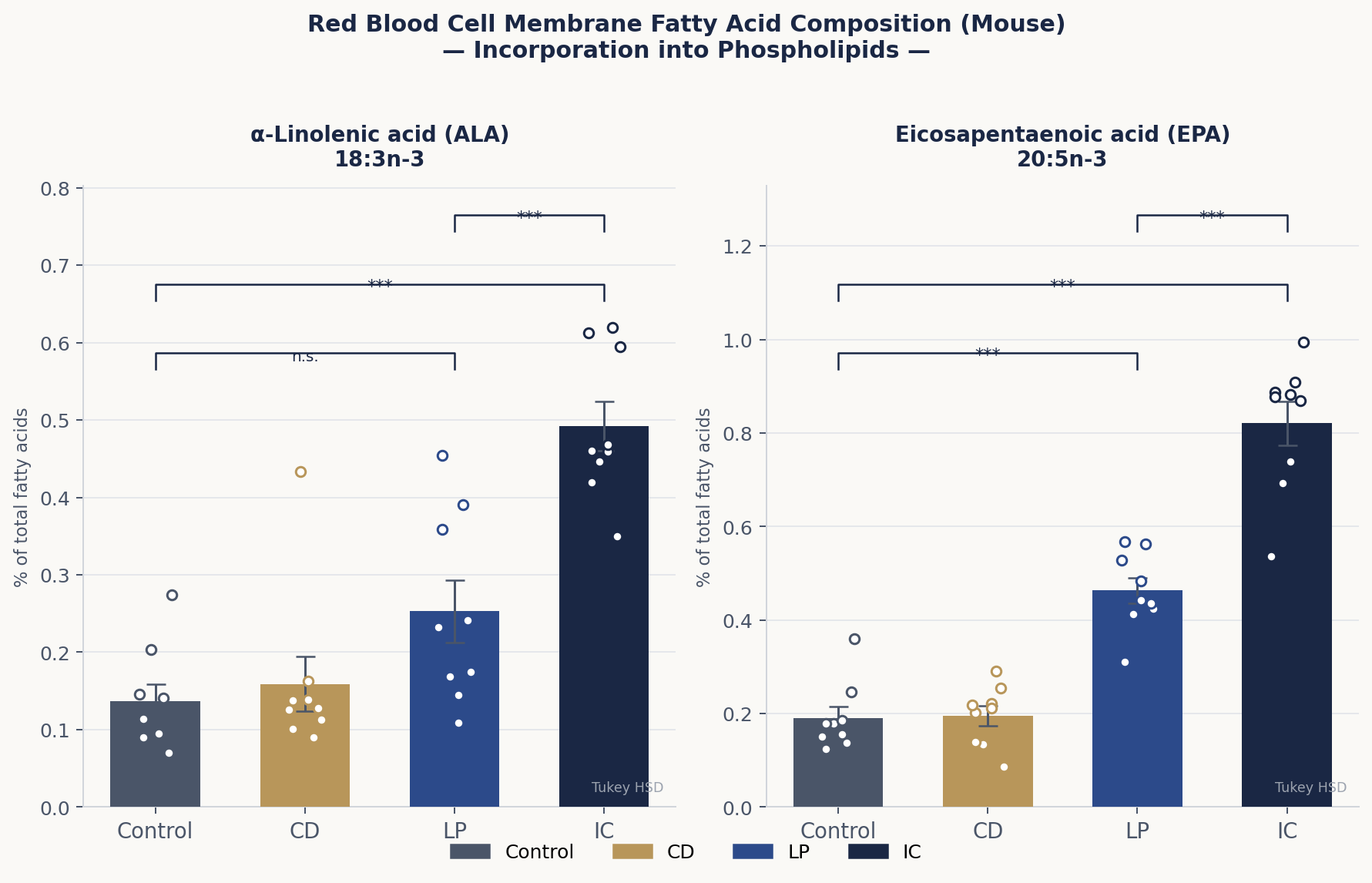
- Red blood cell membranes (utilization as phospholipids): EPA differed significantly among all four groups — Control, CD, LP, and IC (p<0.001). The IC group showed the highest value, indicating that powdered perilla oil further promotes qualitative lipid improvement at the level of peripheral tissues.
Figures & Data
Future Directions
We aim to clarify the detailed mechanisms behind why co-ingestion of γ-CD and perilla oil alters whole-body fatty acid composition — including effects on intestinal digestion and absorption processes and on gene expression. We will continue this research toward creating new food functionalities that efficiently deliver dietary omega-3 fatty acids throughout the body.
Related Publications
Yoshikiyo et al. (2025) International Journal of Molecular Sciences — doi:10.3390/ijms26167776
Yoshikiyo et al. (2025) Molecules — doi:10.3390/molecules30020281
Yoshikiyo et al. (2023) Food Hydrocolloids for Health — doi:10.1016/j.fhfh.2023.100116